Sunday, February 13, 2011

Corynebacterium parvum ANOTHER BACTERIAL "Vaccine" cancer treatment


www.ncbi.nlm.nih.gov › Journal List › Thorax › v.38(1); Jan 1983
http://www.ncbi.nlm.nih.gov/pmc/articles/PMC459478/

http://onctalk.com/wp-content/uploads/2007/03/loculated-pleural-effusion-on-ct.jpg

A malignant pleural effusion can occur as a complication of lung cancer.

A pilot study of topical (intrapleural) treatment with Corynebacterium parvum was carried out in 10 patients with malignant pleural effusions complicating primary or secondary neoplasms and necessitating frequent thoracocentesis for symptomatic relief. The method was aspiration of all intrapleural fluid except a small portion left for dilution, and then injection of 7 mg of a preparation of Corynebacterium parvum suspended in 20 ml of normal saline solution. The treatment was repeated in each case as clinical conditions called for further thoracocentesis. In eight of these 10 patients the treatment resulted in prompt reduction of the rate of accumulation of pleural fluid and a striking change of cell sediment composition, with appreciable reduction in or complete disappearance of malignant cells and a rise in lymphocyte and neutrophil polymorph counts. The best responders were patients with primary pleural mesothelioma. Clinical improvement was evident in all responders.

FULL TEXT
http://www.ncbi.nlm.nih.gov/pmc/articles/PMC459478/pdf/thorax00205-0031.pdf


A pleural effusion is defined as an abnormal amount of fluid in the space between the layers of tissue (the pleura) that line the lungs. If cancer cells are present in this fluid, it is called a malignant (cancerous) pleural effusion.
Causes of a Malignant Pleural Effusion
Almost any type of cancer can cause a pleural effusion if it is present in or spreads (metastasizes) to the chest area. The most common are those mentioned above.
Symptoms of a Malignant Pleural Effusion
The symptoms of a malignant pleural effusion can be very uncomfortable and may include shortness of breath, coughing, and chest pain.

Diagnosis of a Malignant Pleural Effusion

It is important to make an accurate diagnosis of a malignant pleural effusion, since the prognosis and treatment are much different than for non-malignant (benign) pleural effusions. Even with cancer, up to 50% of pleural effusions are benign.

A malignant pleural effusion is often first suspected because of symptoms or findings seen on a chest x-ray or CT scan. If your doctor suspects a malignant pleural effusion, the next step is usually a thoracentesis, a procedure in which a needle is inserted into the pleural space to get a sample of the fluid. This fluid is then examined under a microscope (cytology exam of pleural fluid) to see if cancer cells are present.

If a thoracentesis cannot be done, or if the results are inconclusive, further procedures may need to be done to get an accurate diagnosis. In some cases, a thoracoscopy (a procedure in which a thorascope is inserted into the chest) may need to be done to obtain a biopsy to diagnose a malignant pleural effusion.

Treatment of a Malignant Pleural Effusion


The goal in treating a malignant pleural effusion is palliative, that is, to improve quality of life and reduce symptoms but not to cure the cancer.

If a malignant pleural effusion is very small, it can sometimes be left alone. Thoracentesis can be performed to remove the fluid, but it frequently returns. To prevent fluid from returning, a procedure called a pleurodesis may be done. In this procedure, a chemical such as talc is inserted between the 2 layers of the pleura so that they stick together, preventing fluid from accumulating. This is successful for 60 to 90% of people.

If a malignant pleural effusion persists, surgery may be done to drain the fluid into the abdomen, or a pleurectomy (a procedure that removes part of the pleura) may be performed. New treatments (such as medical pleuroscopy) are emerging to treat malignant pleural effusions as well. Chemotherapy may help with malignant pleural effusions due to small cell lung cancer, but it does not seem to help with those due to non-small cell lung cancer.

Prognosis of Lung Cancer With a Malignant Pleural Effusion

Sadly, the average life expectancy for lung cancer with a malignant pleural effusion is less than 6 months. The median survival time (the time at which 50% of people have died and 50% are still living) is 4 months.


http://www.nature.com/nature/journal/v257/n5525/abs/257396a0.html

Nature 257, 396 - 398 (02 October 1975); doi:10.1038/257396a0

Suppression of cell-mediated tumour immunity by Corynebacterium parvum

HOLGER KIRCHNER, MOSHE GLASER & RONALD B. HERBERMAN

Laboratory of Immunodiagnosis, National Cancer Institute, National Institutes of Health, Bethesda, Maryland 20014

THE use of immunoadjuvants in tumour therapy is based largely on the concept that an increased level of specific immunity against tumour-associated antigens may be achieved by nonspecific stimulation of the immune system (for review see ref. 1). Among these adjuvants, Corynebacterium parvum has received much attention since it represents one of the most powerful stimulants of the reticuloendothelial system in mice2, and is effective in inhibiting tumour growth in several animal tumour systems (for example, see refs 3-6). The mechanisms by which C. parvum interferes with tumour growth have not been established and the tumour protective effect of C. parvum has not been entirely consistent in all systems studied. An immunosuppressive effect of C. parvum has been demonstrated too. In mice, the in vitro lymphoproliferative responses to mitogens and to alloantigens were depressed after injection of C. parvum. It remains to be determined, however, whether a defect measured by these general tests of T lymphocytes may also represent an indication of depressed cell-mediated immunity against tumour cells.

------------------

References
1. Hersh, M., Gutterman, J. U., and Mavligit, G., Immunotherapy of Cancer in Man (Thomas, Springfield, Illinois, 1973).
2. Halpern, B. N., et al., J. Reticuloendothel. Soc., 1, 77--96 (1964).
3. Halpern, B. N., Biozzi, G., Stiffel, C., and Mouton, D., Nature, 212, 853--4 (1966).
4. Woodruff, M. F. A., and Boak, J. L., Br. J. Cancer, 20, 345--55 (1966).
5. Smith, S. E., and Scott, M. T., Br. J. Cancer, 26, 361--67 (1972).
6. Scott, M. T., J. natn. Cancer Inst., 53, 855--65 (1974).
7. Scott, M. T., Cell. Immun., 5, 459--479 (1972).
8. Plata, F., Cerottini, J. C., and Brunner, K. T., Eur. J. Immun., 5, 227--33 (1975).
9. Lavrin, D. H., Nunn, M., Soares, N., and Herberman, R. B., J. natn. Cancer Inst., 51, 1497--508 (1973).
10. Herberman, R. B., et al., J. natn. Cancer Inst., 53, 1103--11 (1974).
11. Kirchner, H., Chused, T. M., Herberman, R. B., Holden, H. T., and Lavrin, D. H., J. exp. Med., 139, 1473--87 (1974).
12. Holden, H. T., Kirchner, H., and Herberman, R. B., J. Immun. (in the press).
13. Kirchner, H., Muchmore, A. V., Chused, T. M., Holden, H. T., and Herberman, R. B., J. Immun., 114, 206--10 (1975).
14. Olivotto, M., and Bomford, R., Int. J. Cancer, 13, 478--88 (1974).

keywords: immune system immunoresponse coley's toxins, bcg, fever treatement

chinese herb - strengthens immune systems


http://en.wikipedia.org/wiki/Astragalus_propinquus

huáng qí (yellow leader) (simplified Chinese: 黄芪; traditional Chinese: 黃芪)

or běi qí (traditional Chinese: 北芪), huáng hua huáng qí (Chinese: 黄花黄耆)

A. propinquus is used in traditional Chinese medicine, where it is used to speed healing and treat diabetes.[4]  In western herbal medicine, Astragalus is primarily considered a tonic for enhancing metabolism and digestion and is consumed as a tea or soup made from the (usually dried) roots of the plant, often in combination with other medicinal herbs. It is also traditionally used to strengthen the immune system and in the healing of wounds [5]  and injuries.[6]  Extracts of A. propinquus are used in Australia as part of a commercially available pharmaceutical MC-S to stimulate production of peripheral blood lymphocytes.

A. propinquus has been asserted to be a tonic that can improve the functioning of the lungs, adrenal glands and the gastrointestinal tract, increase metabolism, sweating, promote healing and reduce fatigue.[7] A mix of Astragali Radix and Salviae Radix, has been used to treat patients with chronic fatigue.[8]

There is a report in the Journal of Ethnopharmacology that Astragalus membranaceus can show "immunomodulating and immunorestorative effects.",[9]The herbal extract supplementation in drinking water can induce an immune stimulation response in immunosuppressed chickens[10] It has been shown to increase the production of interferon and to activate immune cells such as macrophages.[3]

There is a report in the journal Rejuvenation Research titled A Natural Product Telomerase Activator As Part of a Health Maintenance Program which shows that an extract of Astragalus propinquus, called TA-65®, can activate Telomerase extending the lengths of the shortest Telomers which protect the terminal DNA at the ends of all chromosomes. Telomer loss is associated with errors in cell division and is thought to be the primary cause of aging.

The active constituents of the roots (Radix Astragali) include polysaccharides, triterpenoids (astragalosides) [11] as well as isoflavones (including calycosin and formononetin) along with their glycosides and malonates.[

Other Astragalus spp. are known to cause severe poisonings in livestock due to indolizidine alkaloids, aliphatic nitro compounds and accumulated selenium.[14]  None of these constituents have been detected in the medicinal species Astragalus membranaceus used in dietary supplements and TCM preparations.[15]


Saturday, February 12, 2011

deficiency of fever = high risk of cancer = prophylactic fever

http://wwwdelivery.superstock.com/WI/223/1663/PreviewComp/SuperStock_1663R-24176.jpghttp://picturesofthermometers.com/images/ear_thermometer.jpg

The case for prophylactic fever treatment (every year).


Fever is one of the pathophysiological symptoms accompanying the response of the host to infection and inflammation.
Although the adaptive value of the fever response has been well documented in laboratory studies, its role in clinical
medicine is still under debate.

Two of the hypotheses studied state that endogenous mediators of fever may be involved
in establishing the Th1 immunologic phenotype of the host and in improving general immunologic surveillance.
It is known that these factors play a significant role in defence against tumour cells. Therefore, in the present study
we tested the hypothesis that patients diagnosed with cancer reveal a history of fewer fevers during the disease
than control, healthy volunteers.

18 questions were asked concerning the history of fever prior to diagnosis from
355 persons suffering from cancer, and 244 healthy controls, matched for age and living in Poland. Cancer patients
reported a lower incidence of fever during illness than controls. The percentage of cancer patients and controls
who reported no fever during infections was 83.10% and 56.97%, respectively.

Similarly, 16.90% of cancer patients
and 43.03% of controls reported always experiencing fever during infections. The results of our study support the
hypothesis that during their lifetime cancer patients experience less fever during infection than healthy controls.
Key words: fever, infection, cancer, allergy, spontaneous regression

INTRODUCTION

Fever is a specific and well-coordinated pathophysiological
phenomenon associated with infections and trauma, and
manifested by an increase of body core temperature above
normal. It is a part of an acute phase response (APR) - an early
inflammatory response consisting of a host of immunologic,
endocrinologic and neurologic alterations. APR results in
metabolic and behavioural changes collectively called 'sickness
behaviour' [1, 2]. Thus, fever has a significant diagnostic
value which, however, is mostly regarded as an unpleasant
and preventable weakening phase of disorder. In spite of the
overwhelming antipyretic therapy and widespread use of the
fever-preventing drugs, there is ample evidence demonstrating
that fever correlates with increased survival and better
prognosis during microbial infections [3, 4, 5].
Studies into the mechanism and phylogeny of fever indicate
that fever evolved as a constituent of the innate immune
response [4, 6]. Thus, from the biological point of view, fever
is an adaptive response in interactions between the host
and the pathogenic microorganisms. The key steps of the
mechanism of fever have been thoroughly investigated during
the last decades. Briefly, fever is triggered by microbial factors
and products known as 'exogenous pyrogens' or 'pathogen
associated molecular patterns' (PAMPs). Structures such as
lipopolysaccharides (LPS), peptidoglycans, porin complexes,
lipoteichoic acid, lipoarabinomannans, bacterial DNA,
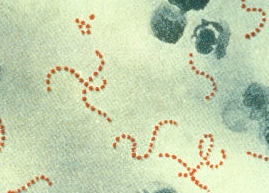
mycoplasma lipoproteins, staphylococcal and streptococcal
proteinaceous superantigens constitute a major group of the
pyrogens of gram-negative, gram-positive and mycobacterial
origin [7-12]. In response to the PAMPs, the immune cells
of an infected organism generate a host of mediators called
'endogenous pyrogens'. Among them are cytokines such as
IL-1, IL-6 and TNF-á [14]. Endogenous pyrogens stimulate
the production of prostaglandins of the E series (PGE2) which,
in turn, act on the fever-mediating thermoregulatory region
of the preoptic area of the anterior hypothalamus to shift
upward a thermoregulatory set-point. The presented scheme of
molecular events leads to the stimulation of thermoregulatory
effectors to gain body heat and to drive body core temperature.

Although the net benefit of elevated temperature is still under
debate, it has been demonstrated that fever stimulates number
of key mechanisms of the defence against infections; among
others, it stimulates T cells proliferation and differentiation,
B cells proliferation and the production of antibodies, secretion
of interferons, phagocytosis, and the migration of macrophages
and neutrophils [15, 16, 5].

Despite the well documented ubiquity of fever, there are
clinical reports suggesting a decreased frequency of fever, or
even the lack of capability of generating fever within certain
groups of patients. Fewer fevers have long been recognized,
especially amongst cancer patients. As early as 1855, the
English surgeon John Laurence acknowledged the fact that
cancer patients have a "remarkable disease-free history" [17].

Since then, clinical oncologists have often reported that in
their history cancer patients stressed that they were almost
never ill, and had never been feverish before the onset of
cancer. Consequently, it has been postulated that a prolonged
lack of fever can be considered as a threat of cancer [18]. Also,
the more recent studies of Witzel (1970), Newhouse et al.
(1977), Remy et al. (1983), Grufferman et al. (1982), Ronne
(1985), Van Stensel-Moll et al. (1986), Grossarth-Maticek et
al. (1987), Abel et al. (1991), and Kolmel et al. (1992) [19-27],
among others, have supported the conclusion that deficiency
of fever in the medical history of the patient corresponds
with high risk of cancer. In the present paper we also report a
lower frequency of fever in the population of tumour patients
compared to healthy volunteers.

MATERIALS AND METHODS

The study was conducted during a relatively short period,
from January 2005 - June 2006. Retrospective information
on fever and fever disorders was obtained from 355 cancer
patients and 244 healthy volunteers by use of a questionnaire.
To collect the information we cooperated with the following
health care institutions: The Polish Amazons Club, The
House of Social Assistance in Toruñ, and the Academy for
Fighting with Cancer and Public Hospital in Inowroclaw.

Each patient had a documented tissue diagnosis of cancer
from pathology records. The majority of the examined patients
were from the Kujawsko-Pomorskie province, and the healthy
control volunteers were randomly selected from the same
area. The respondent cohort consisted of 350 women and
249 men. The average age was 52 (ranging from 16-96 years
old) and 57 (ranging from 17-95 years old) for women and
men, respectively. All participants signed the consent for
taking part in the questionnaire studies. Only completely
fi lled in questionnaire forms were analyzed. The collected
forms were entered into the Access Database for evaluation and
statistical analyses. The chi-square test was used to compare
rates of occurrence between patients with cancer and healthy
people.

RESULTS AND DISCUSSION

Nowadays, it is well documented that fever directly activates
defence against various dangers (including cancer cells) [28,
29]. It is also well known that various microbial stimuli are
necessary for the normal maturation of the immune system
[30]. This discovery places in an unfavourable light the
situation of cancer patients, who very often stress that before
diagnosis they could be considerate as examples of health.
They had never been ill, and even if they had, they almost
never been feverish. Moreover, the observation that cancer
patients who experienced a feverish period after surgery
survived significantly longer than patients without fever,
and the fact that spontaneous tumour remission was observed
mostly after a fever, confirms the significant meaning of this
mechanism for a patient's recovery [31]. For this reason we
performed an epidemiological study. Our aim was to discover
whether there is a difference in the frequency of fever episodes
between healthy people and cancer patients, and to check
peoples' attitude to fever. We compared 355 forms fi lled out
by cancer patients, with another 244 forms from healthy
people (Table 1).

It was observed that the frequency of feverish events during a
whole life significantly differed between the 2 groups: 83.1% of
cancer patients, compared to 56.97% of control group, declared
that they never or almost never have been feverish (Table 1,
Fig. 1). Among the cancer patients only 16.9% did recall of
getting fever in compare to 43.03% of control people. This
data are in accordance with Engel's results, who compared 300
cancer patients with 300 patients not suffering from cancer.
People who had never experienced febrile infectious disease
were 46 times more likely to have developed cancer than those
who had had febrile infections [32]. More recent studies also
confirm these earlier results. In 1987, Grossarth-Maticek et.
al., after questioning 1,353 people, indicated that: "episodes
of high fever during the entire life span in the case of an acute
illness as a typical reaction are inversely related to later cancer
incidence" [25]. In 1991, Abel et al., in a case-control study
with 255 cancer patients compared with 230 controls, showed
that patients who had the highest risk for cancer were those
with a low "infectious index" [33]. Kolmel demonstrated the
essential meaning of the number of febrile illnesses, their
length and level concerning the risk of melanoma incidence.
The undergoing of a minimum of 3 fevers above 38.5 °C
decreased the risk of melanoma incidence by approximately
40% [34].

The surprising result of our research was that above 56% of
healthy volunteers have never been feverish (Fig. 1). During 17
months of our research we found a few cases from the control
group who were subsequently diagnosed as cancer patients.
It is possible that such cases were or will be more frequent.
This could be the cause of erasing the difference between two
groups of our responders.

We observed that there were no significant differences
between cancer patients and healthy control volunteers in
the highest temperature value they recalled having during
their lives. The average, highest temperature during whole life
for the control group was 39.5 C ± 0.8, and for cancer patients
39.2 C ± 0.9. We conclude that this part of cancer patients
who had been feverish (16.9% of cancer patients) can develop
the same level of fever as healthy people. This suggests that
carcinogenesis of some cancers may be indirectly connected
with inefficient generation of fever.

There exist epidemiological studies supporting the
hypothesis that there is an association between febrile
infectious childhood diseases and subsequent cancer risk.
Kolmel et al. demonstrated an inverse relation between the
number of children's febrile infections and the incidence of
malignant melanoma in 271 controls versus 139 melanoma
patients [27]. Exposures to such infections were also associated
with a reduced risk for ovarian cancer [35, 20] and multiple
cancers combined [33, 36]. We investigated whether the
assertion that children's contagious illnesses have a preventive
eff ect on cancer is true or not. After our respondent cohort
examination, significant differences were observed in the
incidences of children's contagious illnesses such as: mumps,
rubella and chicken pox (Tab. 2, Fig.3).

The study by Hoff man et al. suggests that chickenpox and
mumps were associated with an increased risk of cancer [37].
We demonstrate that healthy volunteers suffered from such
disease more often than cancer patients. Our results are in
accordance with Newhouse data who found lower incidence
of mumps, measles and rubella in the cancer group [20].
Hoffman et al. suggested that no significant reduced risk
is seen between cancer and rubella [37], whereas our data
and data by Newhouse showed a lower frequency of rubella
for cancer patients, compared to healthy people. Only data
concerning measles are in accordance - we did not observe any
statistically significant difference in the mortality between
cancer patients and healthy people. We conclude that because
of divergent results, no final statement on the association
between childhood disease and cancer may be made. However,
taking into consideration our results and published data we
must stress that febrile contagious illnesses during early life
are probably not sufficient to protect against cancer because
many of our cancer patients had suffered from such diseases.

We can therefore suppose that not only infection is important
for the stimulation of the immune system against cancer.

We would like to emphasize that infection connected with
fever, which occurs directly before or at the beginning of cell
transformation, is probably of the greatest significance. Our
stance is in accordance with results of Kolmel et al. who also
stressed that febrile infectious childhood diseases were less
protective against cancer than adult febrile infections [27].

It is known that endogenous mediators of fever play a
significant role in defence against tumour cells [38, 39]. We
hypothesize that these factors may be involved in establishing
the Th1 immunologic phenotype of the host. We suppose
that cancer patients who had never been feverish, prefer Th2
phenotype. Similarly, allergy is an immunological disorder
with a predominant Th2 inflammatory response [40]. The
debate about the relationship between allergy and cancer is
not recent [41]. The "hygiene hypothesis" proposes that lack
of early life infections may up-regulate allergic disorders [42].

We found an appreciable difference in the incidence of allergy
between cancer patients and healthy volunteers (Table 1,
Fig. 4). If a decrease in the number of infections is essential
for allergic disorders and for cancer, it is surprising that only
5.92% of cancer patients, compared to 22.13% of the control
group, suffered from allergy. Moreover we have data which
suggest that cancer and allergy exclude one another (data not
published). The question whether allergy really is a protective
factor for cancer, remains unanswered. However, efficient
redirecting the Th2 response in favour of Th1 will probably
be most essential for both disorders.

The second part of our questionnaire form contained
questions to check people's general attitude to fever (Table 1).
We discovered that people regard the 2 notions -- fever and
illness -- as being identical. A small rise of temperature
from 36.6 °C to 37.5 °C by both groups was treated as an
uncomfortable symptom (Table 1, Fig. 5). The temperature
regarded as fever by cancer patients was 37.82 °C ± 0.4; by
control 37.83 °C± 0.5. People tried to eliminate this disparate
state, in spite of the fact that it is a symptom which informs the
body of the danger related to infections. As mentioned earlier,
on the one hand fever may directly debilitate the pathogen, on
the other hand it induces a cascade of host defence mechanism
that increases the action of the immune system [15, 16].
The rise in body temperature is closely related to the ability
to increase cyclooxygenase (Cox) products of arachidonic
acid (especially prostaglandin PGE2). There are actually 2
Cox enzymes -- Cox-1 and Cox-2 -- both of which produce
prostaglandins that promote inflammation, pain, and fever.

Nonsteroidal anti-inflammatory drugs (NSAIDs) block the
Cox enzymes and reduce prostaglandins throughout the
body. In consequence, ongoing inflammation, pain, and
fever are reduced [43]. Because of easy access, nonsteroidal
anti-inflammatory drugs are very popular. Nowadays almost
everybody can reduce or block fever. The necessity for using
medicines against fever because of the rise in body temperature
was declared by 91.4% of people in the control group, and
87.8% of the cancer patients (Fig. 6). We found that 67.96%
of the control group and 74.6% of cancer patients take these
medications before their temperature reaches 38 °C (Table 1).

Moreover, 60.89% of the control group and 51.42% of cancer
patients take medicines against fever always, or almost always,
even after a small rise in body temperature (Fig. 7). For this
reason we can suppose that episodes of really high and long-
lasting fever actually do not happen. There are a number of
prospective and retrospective studies indicating that febrile
infections lower the risk of cancer, and can be associated
with the spontaneous remission of various tumours [44, 28].

Early use of NSAIDs may deprive us of this chance. In 1998,
Mastrangelo et al. revealed that a reduction of infections in
the second half of 20th century caused an increase in cancer
cases. They discovered that a 2% decrease of febrile illnesses
in one year correlated with a 2% increase in tumors after
10 years [45]. This reduction of infections is undoubtedly
connected with so-called "increase in life hygiene" and with
the use of NSAIDs and antibiotics. Taking into consideration
the influence of fever on the immune system, we consider that
the use of NSAIDs should be more prudent.

Fever is a very important mechanism that supports our
immunological system. Some disorders (cancer, allergy)
seem to be preceded by a lack of fever. Our study confirmed
this notion, despite the fact that it was performed during a
relative short period (17 months). Whether this lack of fever
starts before carcinogenesis or is a consequence of a longterm
process which leads to cell transformation, remains
unresolved. Moreover, we found that more than 72% of all
respondents had never been feverish. It is an open question
whether or not the rare fever episodes evolved naturally, or
are the results of frequent switching off of this mechanism
using NSAIDs which, as we established, people often take
unquestioningly. It is possible that with the passing of time
the immune system ceases developing fever at all and we would
be totally dependent on medicine.

The increase of life hygiene and using NSAIDs is also
connected with the high incidence of allergy. In our study,
we discovered that only small part of cancer patients who
had never been ill and had never been feverish, suffer from
allergy, even though some data suggest that infections are
very essential for protection against cancer, as well as allergy.
This problem should be meticulously examined. It is possible
that artificial induction of fever will be helpful in therapies
against both disorders, and research on this subject haa already
been started [46, 47]. However, nowadays we do not have an
answer to the question: which part of the mechanism of fever
is involved in decreasing risk of cancer and allergy, and which
part participates in the induction of tumour prevention and
remission.

FULL TEXT WITH GRAPHICS by S Wrotek - 2009
http://www.jpccr.eu/archive_pdf/2009_vol_3_nr_1/jpccr_05_str_31_35_Wrotek_et_al.pdf


... sauna drives up the body temperature. It is actually a prophylactic fever ...


http://www.portlandmercury.com/images/blogimages/2008/11/10/r_1226366283_uglycat.jpg
(the world's ugliest cat)

Biological basis of the behavior of sick animals

Benjamin L. Hart

Department of Physiological Sciences, School of Veterinary Medicine University of California, Davis, CA 95616, USA

Received 1 February 1988. 
Available online 18 October 2005.

The most commonly recognized behavioral patterns of animals and people at the onset of febrile infectious diseases are lethargy, depression, anorexia, and reduction in grooming. Findings from recent lines of research are reviewed to formulate the perspective that the behavior of sick animals and people is not a maladaptive response or the effect of debilitation, but rather an organized, evolved behavioral strategy to facilitate the role of fever in combating viral and bacterial infections. The sick individual is viewed as being at a life or death juncture and its behavior is an all-out effort to overcome the disease.

Finally the notion of a prophylactic fever foranimals such as ...... A modest prophylactic fever during the activeperiod may have some ...


Thursday, February 10, 2011

AUDIO mp3 Cancer and the Immune System MacADAM seordy coley

More than 100 years ago, a New York surgeon named William Coley developed an
unusual cancer treatment that produced a high fever in his patients.
Although the treatment was unconventional, it turned out to be successful
surprisingly often. Should Coley's treatment be revived today? How else
might we jump-start the immune system? Find out about the hope for vaccine
treatments for cancer, as well as the status of Coley's fluid. This is the
second in a three-part series on unconventional approaches to cancer.

Guests: Don MacAdam, CEO of MBVax Bioscience in Ancaster, Ontario, Canada.
Online at MBvax.com <http://www.mbvax.com/>

Jonathan Serody, MD, Elizabeth Thomas Professor of Medicine, Microbiology
and Immunology in the Program in Stem Cell Transplantation. He is Director
of the LCCC Program in Malignant Hematology and Co-leader of the LCCC
Immunology Program at the University of North Carolina at Chapel Hill. The
photo is of Dr. Serody.

Elite Tabloid Story - Coley Dashel & Rockefeller

Web-logged from the New York Times (tabloid)

"LAUDABLE PUS"

"The first hope of therapeutic success comes with the observation of the efficiency of unaided nature to accomplish cure....
These cases, rare though they be, are the sum of our hope."
--Pearce A. Gould, "The Bradshaw Lecture on Cancer," 1910

There survives from the beginning of this century an intriguing bit of paper ephemera, notable less for the celebrated names marked down upon it than for a medical future it unknowingly foretells. As the year 1903 drew to a close, a rather well known New York businessman went to a Manhattan doctor of his acquaintance for a routine blood test, which was sent for analysis to an increasingly prominent pathologist at the Cornell University Medical College. The results of these tests, typed out on a half-sheet of paper and dated December 7, 1903, are quaintly minimalist and read as follows:

    Hb 95%
    Red cells 4496000
    Leucocytes 5000
    Mononuclear 44%
    Polynuclear 55%
    Eosinophile 1%

"In the stained specimens," the pathologist noted, "I find nothing abnormal."

There is nothing abnormal about the blood chemistry, either. In quick translation, the results indicate a healthy number of red blood cells, the cells that carry oxygen to the tissues; a low but acceptable number of white blood cells (or leukocytes), which are now known to comprise the main cellular components of immune activity; and a reasonable proportion of the various cells that make up the "white blood." What at first seems so remarkable about this lab report is the confluence of names, interests, and destinies attached to it. The pathological analysis came from James Ewing, who would go on to head Memorial Hospital in New York and become one of the most influential cancer authorities of the twentieth century. The doctor ordering the tests was William B. Coley, who in the early days of the century claimed to have cured a number of inoperable cancer patients with a crude antitumor vaccine. And the blood itself belonged to John D. Rockefeller, even then in the process of establishing the great medical institute that would bear his name and whose philanthropic largesse advanced the interests and causes, to greater or lesser extent, of the other two men whose names appear on this otherwise forgettable bit of biochemical trivia.

All three men were dedicated, in their own way, to a cure for cancer, little realizing that certain clues lay in the very cells whose mundane numbers were dutifully recorded in the blood report. And it bespeaks the twists and turns of nearly a hundred years of research into cancer and immunology that the most important word on that fragile piece of paper is not the celebrated names that have come to symbolize wealth or medical authority but rather the word blood. All of Mr. Rockefeller's millions, all of Dr. Ewing's encyclopedic knowledge, and all of Dr. Coley's clinical intuition could not together purchase the knowledge we have painstakingly acquired in a century of research into the remarkable qualities of blood--its leukocytes and other cells, its proteins, its molecules and factors then unimaginable--all of which are now understood to function, like sections of an orchestra, in the daily biological symphony that is the immune system. In telling the story of how much of that knowledge has come to be won in the treatment of patients, it is most appropriate to begin with that crucial intersection of Coley and the Rockefellers and a disease whose diagnosis, then even more than now, was tantamount to a death sentence: cancer.

The destinies of the Rockefellers and Dr. Coley first crossed more than a decade earlier in a tragedy that, as the tabloids might put it (and the New york times tabloid does put it) , left one young woman dead and the lives of two men changed forever. It began in the fall of 1890, shortly after twenty-eight-year-old William Coley had finished his residency as a surgeon at New York Hospital and entered private practice. He was still a green and impressionable physician, still learning his trade, when a young woman named Elizabeth Dashiell came to see him, complaining about a nagging pain in her hand.

She was seventeen at the time, thoughtful and self-possessed, with a face so firmly etched with determination and good humor that she appears in surviving photographs almost preternaturally poised and mature. Born in Minneapolis, Dashiell had moved with her family to Lakewood, New Jersey, when she was two years old; her father, a minister named Mason Dashiell, had died when she was very young. She might have been one more young woman from small-town New Jersey save for the fact that she claimed as a close friend and confidant one of the most famous, and upon his majority one of the wealthiest, Americans ever produced by this nation: John D. Rockefeller Jr. Painfully shy and awkward as a youth, the customary solitudes and discomforts of adolescence no doubt heightened by the attention drawn by his family's notoriety, the only son of the founder of Standard Oil befriended Bessie Dashiell through her older brother Lefferts, who attended the same private school in New York as Rockefeller; one of Rockefeller's three sisters was also named Bessie, and he came to think of Bessie Dashiell as his "adopted sister." They took carriage rides together, rode horses along the Hudson Palisades, and exchanged long, thoughtful letters. There survives a formal portrait of the two of them: "Johnny Rock," as he would later be known to classmates at Brown University, looking less granitic than Fauntleresque, a soft-featured, frail-looking tulip of a youth, all four buttons of his pinstriped suit firmly fixed, a derby in his gloved right hand, and Bessie Dashiell, seeming to tower over him though she sits to his left, clear of eye and with a slight, knowing smile, gussied up in an overcoat with a fur collar, firm of jaw and beguiling in her self-aware sense of humor. That the stars brought this young woman and her notable friend into the orbit of Dr. William Coley ultimately had a profound effect on American philanthropy and cancer research, to say nothing of Coley's career.

Dashiell had a taste for daring. In the summer of 1890, she had undertaken what in retrospect qualifies as Victorian America's equivalent of adventure travel--a railroad trip across the continent, followed by a jaunt up to Alaska. During the trip, as she later wrote young Rockefeller, she had hurt her hand and thought it had become infected. By the time she had returned to New Jersey in August, the hand was still swollen and painful. As it turns out, the injury stemmed from the most trivial of accidents. Dashiell's hand had at some point become caught and pinched between two seats of the Pullman car in which she was riding. That "slight blow," as her doctor would later describe it, left an ordinary bruise upon the back of her right hand.

After some initial swelling and pain, the injury appeared to improve, but after a week, both pain and swelling grew more severe. To all appearances, it was a typical bruise, and a local physician recommended nothing more than "the usual local applications," probably ice. Nothing about so minor a bump seemed ominous, and hardly anything about this young woman suggested frailty--she had been in excellent health her entire life. Little more than a month later, back in New Jersey, Bessie Dashiell went to her own doctor complaining of continued pain; it seemed more pronounced with motion, she reported. In an attempt to alleviate the mysterious and persistent pain, the hand and wrist were placed in a splint, but that brought only temporary relief. During September, the pain grew so persistent, with occasional sharp and shooting flashes of discomfort, that Dashiell had trouble sleeping. Finally, her family decided to seek medical help in New York City. The man to whom they were referred was William Coley.

Coley, barely a year out of medical school, was already a rising star in New York surgical circles, at a time when surgery itself was the ascendant medical art. He examined her for the first time on October 1, and in the manner of the day, characterized the swollen area according to the scientifically imprecise but useful idiom of the farmer's market; he noted a small, spindly swelling "about the size of half an olive" on the back of her right hand, just above the large joint leading to her small finger. When he pressed down hard to determine if the swelling was mobile, his young patient complained of the pain. He checked for inflammation of the lymph glands under the armpit, which might indicate an infection, and found none.

Several days after this initial consultation, Coley made a more detailed examination. He suspected the problem was caused by a low-grade inflammation of the membrane, or periosteum, covering the bone, a condition known then as subacute periostitis. To make sure, he ventured a closer look; for a shy country boy who'd dithered over the choice of a profession scarcely five years earlier, Coley had quickly learned to wield the knife without conspicuous indecision. Applying cocaine as a local anesthetic, as was common at the time, he made a small incision through the center of the swollen area down to the bone. He noted a few drops of thin pus, but nothing in an amount suggestive of infection. Puzzled, he probed the tender area and observed that the tissue through which he'd cut "seemed abnormally hard and more of a grayish color than normal." He closed the wound with antiseptic dressings and placed the hand once again in a splint.

Coley probably expected to find telltale traces of a discharging infection when he later changed the dressing, but he did not, and when Dashiell reported that the pain and swelling had increased yet again, he must have felt uncertain about what to do next, because the following week he sought the advice of his mentor at New York Hospital, the celebrated surgeon William Bull. Bull, too, thought it was periostitis. He advised "waiting for further developments." Up to this point, Bessie Dashiell may well have viewed the problem as a nuisance, exceedingly painful but hardly more than a lingering, inconvenient bruise. Indeed, her friend John D. Rockefeller may have been more concerned than she; unaware at first that she had been hospitalized, he wrote her a nine-page letter on October 19 detailing his worry ("I cannot tell you how I shall miss my adopted sister on such occasions") and concluding, "I shall hope to receive just a word from you telling where you are and how your hand is doing before many days."

Her hand was not doing well, and sometime toward the end of October Dashiell must have read considerably greater concern in the face of the young surgeon. As her symptoms slowly but steadily worsened, Coley decided on yet a more detailed look. This time he administered ether and expanded his previous incision to expose a three-quarters-inch length of bone. The membrane covering the bone was thick, but the bone itself appeared clean and free of pus, thus seemingly free of infection. The young surgeon scraped away what he referred to as "grayish granulations" and closed the wound once again. Temporary relief ensued for a day or two, but the pain returned "as severe as before" with redness and swelling, and then Dashiell's condition became ever more frightening: she reported losing sensation in several fingers of her right hand. "The pain soon became so severe," Coley remarked, "that I was obliged to give morphine to secure relief."

Clearly, some process other than routine inflammation seemed to be causing the excruciating pain. Coley wracked his neophyte's brain for an answer. There was one other remote possibility to explain what was going on, and Coley reluctantly began to entertain it. The gradual increase in pain, swelling, loss of sensation, and impaired motion, absent an obvious infection or other inflammation, suggested something much more serious: cancer. Specifically, it suggested sarcoma, a malignant disease of the body's connective tissue, such as muscle, bone, and the miscellaneous gristle in between that holds us together (carcinoma, the other main form of cancer, arises in the epithelial, or surface, layer of tissues, and typically affects major organs like the lung, breast, liver, ovaries, and colon). After securing the consent of Bull and Robert F. Weir, chief surgeon at the hospital, Coley obtained a biopsy sample in early November by cutting away a small, wedge-shaped bit of tissue, which was given to a pathologist at the New York Hospital for microscopic examination. On November 6, the worst-case diagnosis came back: the cells on the slide bore the typical signature of cancer, and the pathologist's report read "Round cell sarcoma."

It is not hard to imagine the consternation with which this news might have been received by Bessie Dashiell, if she was told; doctors who practiced nineteenth-century medicine (by which is implied its concealing discretions as well as its often fruitless interventions) were famously circumspect about delivering bad news to patients, to say nothing of delivering it bluntly. In circumstances as dire as these, etiquette hardly mattered. In early July Dashiell had caught her hand between two train seats; now, barely five months later, three of the finest doctors in New York were telling her that, as Coley would later record, "amputation at the middle of the forearm offered the best chance of saving the patient's life." What Coley probably didn't tell her was that even that "best chance" made her odds of surviving at best only about one in ten; and what Coley himself almost certainly did not realize is that his initial exploratory incisions to diagnose the problem may have not only promoted the spread of cancer but accelerated what would become a harrowingly rapid decline, one that would haunt him for many years to come as "one of the most malignant tumors I had ever seen."

Many patients refused such radical treatment, but in this case the family had no choice. On November 8, 1890, little more than a month after he first examined Bessie Dashiell and shortly before her eighteenth birthday, Will Coley amputated her right arm below the elbow.

There is no record of Coley's personal reflections on the operation, whether he considered it a reasonable intervention or thought it a last-ditch, well-intentioned act of barbarism by professionals who had nothing better to offer. His not-quite-matter-of-fact recitation of Dashiell's ensuing decline suggests he expected a better outcome. "The appetite remained very poor and she did not regain strength as rapidly as I had hoped," he admitted to colleagues later.

Three weeks after the operation, Dashiell experienced extreme abdominal pain lasting several days before it went away. There was "no indiscretion in diet," as Coley put it, to account for the sudden pain. A more seasoned physician might have recognized it as a sign that seeds of malignancy, hatched in one part of Dashiell's body, had spread and nestled in other hospitable niches of the body, there to expand and proliferate as satellite malignancies known as metastases. Indeed, like some cruel, time-lapse movie of malignancy, tumors began to sprout up on Bessie Dashiell's body with alarming speed. On December 11, her doctors discovered a small lump, about the size of "a small almond," in her right breast; the following day, two smaller nodules appeared on her left breast where none had been the day before. A week later, the lymph glands under one armpit became swollen and painful. More lumps appeared in the breasts. Severe aches developed in the left thigh, and she experienced "almost complete anorexia," too weak to go out walking by Christmas. "The pain was so severe," Coley noted, "that the patient had to be kept under the influence of opiates."

The course of Dashiell's illness became a grim seminar for Coley on the speed with which an aggressive cancer can sweep through and conquer the human territory. She began to lose sensation in her lower lip and chin, then in some lower teeth. Dull pain returned to her abdomen. By the first of the year came jaundice; now the liver was failing. More small nodules cropped up on her chest and trunk; more lymph nodes hardened. "From this time," Coley noted, "the loss of strength and flesh was very rapid. She could take almost no nourishment, even liquid causing severe pain in the abdomen." Two weeks after Christmas, Coley located by touch a well-defined and enormous tumor occupying the whole of the abdomen above the stomach; he estimated it to be the size of "a child's head." The liver was enlarged, the heart began to fail. Like street sweepers tidying up after a nuclear holocaust, doctors plied her with large amounts of digitalis and as much brandy as she could stomach. By January 20, the end was in sight.

The endgame in cancer is never pretty, less so in an era where doctors chased rather than managed the last ghastly symptoms. The breast tumors had become the size of goose eggs, the abdominal tumor even larger; the length of her body from head to toe was stippled by small tumors that Coley likened to buckshot or split peas. Last came the vomiting, several times a day, though she had had no solid food; soon, she was regurgitating copious amounts of blood. "The attacks occurred almost hourly," Coley noted, "and were very exhausting to the patient in her extremely weak condition." Elizabeth Dashiell remained conscious of this horrific piracy of her eighteen-year-old body until very nearly the end, when finally, mercifully, she died at home in New Jersey at 7 A.M. on January 23, 1891. Coley was at her bedside and signed the death certificate.

How much meaning can such a short and tragic life embrace? John D. Rockefeller Jr., no doubt frantic when he finally learned of the gravity of Dashiell's illness, visited her at home not long before her death and attended the January 25 funeral. Mary Dashiell later sent him a keepsake, a celluloid court-plaster case that her daughter had cherished. "I cannot tell you how deeply I appreciate your kindness in sending me something which belonged to Bessie and which she enjoyed," he wrote in reply. "This token will be a continual reminder of her beautiful life and death, and I shall never look at it without being thrilled with the thought of her patient endurance of such continued suffering, and tender regard for those about her, lest they should be made unhappy or sad by her pain."

His grief had two consequences, one short-term and one of truly visionary sprawl. The shock of Dashiell's death left Rockefeller in no shape to attend college; slated to begin his first term at Yale University during the period following Dashiell's death, he spent the time instead tending to the grounds of Forest Hill, the Rockefeller estate in Cleveland. The raw adolescent wound of losing a beloved soul mate, especially for a boy who probably didn't have many, could only leave a deep and lasting scar, and although many a mourner vows to channel her or his grief into practical good, few in history were as well placed as John D. Rockefeller Jr. to translate such resolve into something of enduring impact, and few were as good to their word. As a young adult, he dedicated much of his philanthropic effort to the conquest of cancer; those efforts began five years after Dashiell's death, in 1896, with dabbling support for William Coley's research (the two men remained friends throughout life), grew prodigiously with his family's creation of the Rockefeller Institute for Medical Research (now Rockefeller University), and led ultimately to a multimillion-dollar gift that allowed creation of Memorial Hospital (now Memorial Sloan-Kettering Cancer Center) at its present site in New York City. Asked many years later how he became interested in cancer research, Rockefeller replied, "I think it goes back to Bessie Dashiell ... Her death came to me as a great shock."

Dashiell's swift death left her physician no less shaken. Only ten years older than Bessie, William Coley was too young, too professionally unseasoned, to shrug off the case. In his very last scientific paper, written nearly half a century later, he reiterated that Dashiell's case made a "deep impression." He felt helpless in the face of Dashiell's illness, and was naive enough to think he could do something about it. The case sobered Coley not only because of the speed with which the cancer killed, but because of the crude, puny, and utterly ineffectual obstacles hurled by her doctors to impede its fatal course. Medicine, as it was practiced then at New York's finest hospitals, had nothing better to offer than morphine, brandy, and the bone saw, and nothing to show for the effort. William Coley appears to have been one of those stoic personalities who betray no emotion, not because he didn't feel any but probably because he felt too much; when he related Dashiell's case history to surgical colleagues several months later at the New York Academy of Medicine, cataloging each decrement of her mortal illness with the detached precision of the keen clinical observer he had already become, he hinted at his own frustration only in his concluding remarks. "A disease that, starting from an insignificant injury, can attack a person in perfect health, in the full vigor of early maturity, and in some insidious, mysterious way, within a few months, destroy life, is surely a subject important enough to demand our best thought and continued study."

Coley's "continued study" began almost immediately. Curious about such a starkly malignant disease, he decided to search the hospital records for past cases of sarcoma to learn more about this generally rare disease. In the course of this research, he stumbled upon an unusual case--an aberration, really--that stopped him in his tracks. And that is how, several months after the death of Elizabeth Dashiell, in the spring of 1891, Coley found himself in the midst of a most unusual epidemiological manhunt on Manhattan's teeming Lower East Side. For several weeks, during the hours after work, he searched the tenement neighborhoods, climbing interminable flights of stairs, inflicting his pidgin German on startled tenement dwellers who opened their doors to an earnest young doctor, certainly not the kind of man regularly seen paying house calls in those precincts. At each door he inquired after a German immigrant, a house painter, last seen at New York Hospital in February 1885. The man's name was Stein, and he probably bore a large, telltale scar behind his left ear.

"Nature often gives us hints to her profoundest secrets," Coley said on another occasion, "and it is possible that she has given us a hint which, if we will but follow, may lead us on to the solution of this difficult problem." Nature had given Coley a hint; now he determined to follow it down whatever avenue, up whatever steps, through whatever door, to find this man Stein; and through Stein to explore the biology of an apparent medical miracle.

Banging on tenement doors may seem like a profligate waste of a surgeon's valuable time, to say nothing of a professional hazard to his valuable hands, but medicine was different at the turn of the century in many respects, and in Coley's case his willingness to augment knife and gown with medical gumshoeing speaks well of his ability to practice a new kind of medicine. If there has been a reluctance to accord him recognition as the father of immunotherapy, it may in part relate to a tendency to hold this thoroughly nineteenth-century physician to twentieth-century scientific standards, and perhaps also to overly extravagant claims made on his behalf by champions of alternative approaches to cancer treatment. In order to appreciate what William Coley accomplished, it is essential to appreciate who he was--and just as important, who he was not. He was a wonderful clinician, a superb surgeon, an esteemed colleague of the major medical workers of his generation, including William Welch of Johns Hopkins, Harvey Cushing of Harvard, and the Mayo brothers. One thing he was not, however, was a scientist; he never trained in any form of laboratory work, and never truly understood the rules by which the increasingly scientific approach to medicine was being played. Yet in straddling two distinct medical eras, he managed to be a modernist and old-fashioned all at once; and in order to understand why Coley's anticancer vaccine received the largely indifferent reaction it did, it is essential to understand Coley as a man ahead of the times and yet behind them, too.

His roots were strictly rural. William Bradley Coley was born on January 12, 1862, in the small Connecticut community of Saugatuck, just north of Westport, in a district that formed part of a heavily wooded, gently rolling sixty-five-acre parcel of land granted to the Coley family by King George III in 1763 and informally known as "Coleytown." His father, Horace Bradley Coley, taught in a one-room schoolhouse and farmed corn, onions, and potatoes to make ends meet while his mother, Clarina Wakeman Coley, tended the house. The family traced its American roots back to Samuel Coole, who arrived in the Massachusetts Bay Colony in 1631. Theirs was a clan of solid (and stolid) citizens: militiamen, selectmen, church deputies, known for their farming, teaching, and upright lifestyles.

An empathetic physician throughout his career, Coley may have acquired the skill involuntarily, beginning at an early age. His immediate family suffered devastating losses to the kinds of common mortal illnesses that would, by the end of Coley's career in medicine, be regarded as novelties. Horace Coley's first wife, Polly Sophia Wakeman, died in 1854, probably of puerperal (childbirth) fever; his second wife, Clarina, mother of William Coley (and sister of the recently deceased Polly), succumbed to typhoid fever when her son was only nine months old. Of Coley's first two stepsisters, one later died of peritonitis, the other of an unknown fever; Coley's sister Carrie, probably the most beloved of all in his immediate family, died in 1892 after a life-threatening pregnancy and an emergency abortion late in the first trimester (which Coley reluctantly performed himself), and Coley also lost his second-born son Malcolm, who died in 1901 at age five of an acute gastrointestinal infection, after less than a week of illness. Following the death of William's Coley's natural mother, Horace Coley remarried a woman named Abbie Augusta Gray; a much-beloved stepmother to William Coley during the crucial years of his upbringing, she too died, in November 1879. By the time Coley turned eighteen, in other words, he had lost two mothers.

By that age, however hardened by the deaths around him, Will Coley had developed into a slender, handsome young man of five feet eight, brown-haired and brown-eyed, of notoriously shy demeanor. Intelligent and deeply religious, he attended a private academy in Westport, where his first exposure to the classics would initiate a lifelong love affair with great literature and account for two of his greatest extracurricular passions: reading and the collection of rare books.

Nothing about the upbringing or education of William Coley advertised the imagination with which he perceived the importance of his toxin therapy or the genial tenacity with which he defended it against many detractors over many decades. When he set off by train to New Haven for his first year at Yale College, having been accepted into the class of 1884, it was like crossing some intellectual Bosphorus and setting foot upon a newly discovered and exotic continent of books, ideas, society, culture--all the more so for someone who, if truth be told, probably reminded no one of a worldly, urbane dandy. As the journal he began to keep suggests, life before Yale was virginal in every respect ("Played cards! I never knew the name or value of a card before but learned so quick," he wrote, in a brief verbal frisson, familiar to all undergraduate initiates to the pleasures of sin, in February 1882; the wonder is that it took until his sophomore year to make the discovery). The language quoted in his diary is of someone just learning that he has a voice, much less testing its limits: painfully earnest and literal, rarely witty, uncomfortable with strong opinions, Coley's journal rarely dares to scratch beneath superficial observation. "Completely carried away with it," he wrote glowingly of Longfellow's poem The Courtship of Miles Standish.

Coley had never played cards before going to college, had never gone to the theater, had never smoked. He found time each week to teach Sunday school, read his Homer and Aeschylus for classes and his Paradise Lost and Samson Agonistes with greedy pleasure during breaks, and supplement his always threadbare finances by ghostwriting compositions for less gifted students while tutoring others in math and physics. He must have been an excellent tutor: he once commanded $32 for several sessions, which sum covered roughly 20 percent of the annual tuition at Yale. He was not "tapped" for membership in a secret society, did not compete in sports, did not join clubs [except the freshman debating society), and decided against attending the Junior Promenade one year because he could not afford the $4 ticket. As for recreation, he tended toward the more economical diversions: ice-skating, boating, most of all walking. As his classmates later recalled, Coley was "very quiet and reserved at Yale--no extracurricular activities and never athletic." Of his personality, all we are told is that he possessed a "merry laugh."

To anyone who has made the acquaintance of that humble and self-effacing creature, the surgeon, Coley's ultimate migration to that branch of medicine seems almost temperamentally precluded by his earnest indecisiveness. His initial exposure to the medical profession came by way of his extended family; when he was home on break from college, he would sometimes accompany his uncle, Dr. Joseph Henry Wakeman, on house calls in the Redding, Connecticut, area in the doctor's horse-drawn buggy. But he was far from decided upon a career in medicine. In January 1884, just a few months short of graduation, he described a fitful night in bed trying to solve the problem: "I lay awake a good part of the night listening to the fierce blast of the wind and rain upon the roof and meditated whether to study the Chemistry Optional next term and take Medicine as a profession or to take Law. I could not come to a satisfactory decision, although I did not give up until nearly morning."

After much fretting and procrastination, he punted--he decided to accept a post as "assistant principal" (a glorified term for an overworked instructor) at the Bishop Scott Grammar School in Portland, Oregon, where he could set aside some money and ponder the decision anew. The school was run by a Yale graduate named Joseph Wood Hill, who happened to be the husband of a distant cousin, Jessie, which fact undoubtedly complicated negotiations when Coley insisted on timely installments of his $600 a year salary, always seemingly in arrears. After taking the Northern Pacific railroad across the continent, he remained two years in Portland, teaching everything from Greek and Latin to mathematics, gazing out the window of his room at Mount Hood, saving close to $750 with which to further his education, and still pondering in which profession to invest it. Here his reading brought him into contact with two influential figures, one medical and the other spiritual.

The latter, interestingly enough, was Giordano Bruno, the radical sixteenth-century Italian humanist. This Renaissance scholar championed the Copernican, sun-centered view of the solar system; believed the universe was infinite and composed of many systems like our solar system; posited an atomic-based concept of matter; spoke of the Bible as a book of moral but not scientific authority; lectured on a peaceable utopia where all religions coexisted in an atmosphere of harmony and dialogue; dabbled, fatefully, in mysticism; and aroused the fury of ecclesiastical authorities by steadfastly sticking to these radical, unorthodox, and clearly erroneous beliefs. Bruno did not, like Galileo, mumble his defiance under his breath to save his skin, for which insolence Bruno was ultimately burned at the stake in Rome's Campo de' Fiori in 1600. Coley filled several pages of his notebook with admiring observations about Giordano Bruno. "Drank deeply of the spirit of the Renaissance," he wrote. "Accepted the discoveries of Copernicus, and used them as a lever to push aside antiquated systems of Philosophy." Coley never thought of himself as a martyr, but the lifelong vicissitudes of winning acceptance for his work might easily have reminded him of Bruno's travails.

Another book exerted a more immediate impact. Coley happened to read the autobiography of J. Marion Sims, the celebrated surgeon who had helped found the New York Cancer Hospital in 1884. "I have been asked many times why I studied medicine," Sims wrote in a passage Coley dutifully copied into his notebook. "There was no premonition of the traits of a doctor in my career as a youngster; but it was simply in this way: At that day and time, the only avenues open to a young man of university education were those of the learned professions.... A graduate of college had either to become a lawyer, go into the church, or to be a doctor.... I would not be a lawyer; I could not be a minister; and there was nothing left for me to do but to be a doctor." Not only did Coley reach the same conclusion, but twenty years later, this Oregon schoolteacher would play a major role in shaping the hospital founded by Sims into the city's--and ultimately the nation's--preeminent cancer research institution, Memorial Hospital.

In September 1886 Coley began his medical education at Harvard; it says something about the somewhat less rigorous standards of medical training in the days prior to Abraham Flexner's historic 1910 report, which urged massive reform in medical training and modernized American medicine, that Coley was allowed to enter the three-year program at Harvard Medical School as a second-year student largely on the strength of spending several months during the summer of 1886 accompanying his uncle on rounds in a horse and buggy. At that time many medical schools were not affiliated with a university, most required only two years of study, and hardly any used a standardized examination to test the competence of would-be practitioners before unleashing them on an increasingly wary public. Harvard was certainly among the best of the lot, but it too was struggling to absorb and incorporate the profound changes in medical science, of which there were many in the latter half of the nineteenth century: Joseph Lister had introduced the concept of treating surgical wounds with antibacterial substances (antisepsis) in 1866, followed soon after by the technique of using sterile conditions (asepsis) in the operating theater, and these concepts remained so new during the period of Coley's education that their merits were still heatedly debated in some American hospitals; Louis Pasteur had proposed the germ theory, thereby implicating infectious microorganisms as the cause of infectious disease; and Robert Koch had, just four years before Coley's matriculation, identified the mycobacterium that caused tuberculosis, a disease then claiming roughly 5 million lives worldwide each year. Medicine was in the throes of the most profound changes since the Renaissance, although not all the changes had sifted down to the schools.

Nonetheless, Coley managed to engineer for himself--and it was quite as much his doing as Harvard's--a remarkably liberal, unusually modern medical education. Whereas the conventional curriculum involved little more than an endless series of lectures on anatomy and physiology, chemistry and pathology, Coley had extraordinary good luck in obtaining on-the-job training. During the summer following his first year of medical school, in 1887, he happened to be visiting two friends from Yale who served on the staff of New York Hospital, and they invited him on the spot to fill in for a doctor who had taken sick leave, which is how--at age twenty-five, still without a medical degree, and at a moment's notice--Coley found himself patrolling the corridors of a major metropolitan hospital as "Acting Junior Surgeon" for six weeks. The title almost certainly exaggerated largely factotum responsibilities, but in an era that had barely begun to embrace the educational wisdom of exposing students to actual clinical situations in the wards of hospitals, Coley found himself not merely exposed but plunged into real doctoring: dressing wounds, handling the "etherizing" at operations (that is, serving as anesthesiologist), observing the modern technique of surgical asepsis, acquiring hands-on experience during a hands-off era of medical education. His very first medical paper discussed heat-related illnesses, based on the many sunstroke victims he treated during the summer of 1887. To his sweetheart he wrote with an apprentice's pride, "I sewed on a finger the other day and expect to amputate one today."

Even Coley could not have realized at first how fortuitous that summer's employment would turn out to be. He not only landed in one of the premier private hospitals in New York, but in fateful proximity to two of the best surgeons in the country. Robert Fulton Weir, described in Walter Graeme Eliot's Portraits of Noted Physicians of New York, 1750-1900 as "one of the three greatest surgeons of the day in N. Y. City," was a wiry, gray-bearded fellow with a hooked nose and sharp, hooded, deep-set, and lively eyes, who in formal portraits appears like someone too restless to dress well, pose well, or let go of a raptorlike intensity that stiffened his face with impatience. In addition to a teaching appointment at the College of Physicians and Surgeons, Weir later served as president of the New York Academy of Medicine. The other was a disciple of Weir's named William Tillinghast Bull. The name conjures up an image of Taftlike bulk and Ashmolean sideburns: Bull was indeed a heavyset young man thirteen years older than Coley, his mustache slicked to pinpoints, hair parted smartly down the middle as befits the ladies' man and befriender of nurses he was said to be, a sharper dresser than Weir, with less delicate hands and chilling, distant eyes. He achieved early celebrity in 1884 when, pioneering a form of emergency surgery at the Chambers Street Hospital that has lamentably become ever more essential to twentieth-century medicine, he is said to have performed the first exploratory operation to treat an abdominal gunshot wound; Bull enjoyed such local celebrity that when he became ill with cancer in 1908, the diagnosis and his subsequent months-long struggle against the disease was front-page news in the New York Times and other papers. His influence on Coley is obvious: the young surgeon's second paper, "Treatment of Penetrating Shot-Wounds of the Abdomen," appeared the following year in the Boston Medical and Surgical Journal, forerunner of the New England Journal of Medicine.

The historian Paul Starr has written of the transformation of American hospitals "from places of dreaded impurity and exiled human wreckage into awesome citadels of science and bureaucratic order," and Coley had the good fortune to enter the field just as the foundation stones of that massive new edifice were set into place; indeed, he later claimed to have entered medicine "at the most opportune time in a thousand years." Weir and Bull, for example, aggressively championed the use of aseptic conditions in the operating theater, and Coley learned the technique at their elbows. With each passing year, medicine became more of a science-driven enterprise. Bacteriology's assault on infectious disease represented the leading salient, and collateral fields like microbiology and immunology moved swiftly through the breaches that breakthroughs against infectious diseases created; aseptic techniques allowed surgeons to treat otherwise minor but problematic conditions like hernias and fractures without fear of mortal infection. And significantly, Coley's career would enact in miniature one of the emerging and most enduring conflicts of twentieth-century medicine, when for the first time the duties of the conscientious physician sometimes worked at cross purposes to the needs of the rigorous researcher. With the rise of "scientific medicine" came a newly hyphenated medical creature, the physician-researcher, who straddled with great difficulty the chasm between bedside and laboratory bench.

With the summer experience under his belt and, a year later, his Harvard education behind him, Coley accepted an invitation to be a surgical "Interne" at New York Hospital, beginning in January 1889 (as a last grand adventure before New York, he signed on in August 1888 as ship's surgeon to the Kennard, a barkentine that plied the human trade bringing cheap immigrant labor from the Azores to the textile mills surrounding Boston). Coley's first year in New York was exhilarating, depressing, determining. He arrived in November 1888 and found an apartment at 18 West Sixteenth Street, just next door to New York Hospital, then located on Sixteenth Street between Fifth and Sixth Avenues.

This venerable institution, founded five years before the Declaration of Independence was signed, had relocated to Sixteenth Street in 1875 and received a handsome face-lift, with a redbrick facade and mansard roof. There were nine house physicians at the hospital, who hustled to attend some 375 patients daily; Coley served until July 1889 as a "junior walker," or intern, graduated to senior walker that summer, and was promoted to house surgeon early in the spring of 1890. His skill in surgery must have been considerable, for in addition to his internship at New York Hospital, William Bull secured appointments for Coley at the Post Graduate Medical School, where he had an instructorship in surgery, and at the Hospital for the Ruptured and Crippled, where he would serve for some forty years, ultimately as chief surgeon (this latter is now the Hospital for Special Surgery, where New York Mets and Jets and other professional athletes take their well-heeled joints and bones for repair). There he championed the use of the "Bassini procedure" to treat hernias, a technique he performed thousands of times during his career. At one point in the midst of this intense apprenticeship, Coley's beloved Alice Lancaster broke off their relationship, though they would ultimately reconcile and marry on June 4, 1891, after several intense months of activity that would forever shape the trajectory of Coley's future years.

With early success came lifelong paradox. The boy who milked cows and plowed the fields of his father's Connecticut farm later ministered as personal physician to a rich dowager on one of her European vacations; the boy who walked six miles to Sunday school at the Methodist church in Easton became the man about town who insisted on a chauffeur, in part because he never learned to drive a car. The teenager who shunned college social events for want of money (and, probably, want of social confidence) would later join fifteen medical societies and ten social clubs, reorganize the Sharon, Connecticut, golf club, and exchange chummy letters with robber barons and their kin. The boy raised in a culture of thrift and deprivation became the successful surgeon who perpetually lived beyond his means, who dabbled in real estate speculation, and whose financial adventurism was so ill-advised that near the end of his life he could barely pay for medical treatment for his dying wife.

Yet here is the most fascinating paradox of all. The man who made quite a good living by use of medicine's crudest utensil, the knife, stumbled upon its subtlest of healing tools: the blood, with its cells and its wondrously potent ensemble of molecules. Because he was never trained as a scientist (and never really became a great one during his lifetime), Coley failed to cast his work into a rigorous, molecular idiom; nor could he have, given the technical limitations of the day. Whatever credit he deserves--and he deserves a great deal--derives from the fact that he was an exceptionally conscientious physician who made careful observations, believed what he had seen, and clung to those beliefs with a tenacity that emulated what he regarded as Charles Darwin's greatest virtue as a scientist: doggedness.

Of all his many qualities, that was perhaps the one that served him best. And he would have a lifetime of controversy to put his doggedness to good use.


Histopathological diagnosis is definitive BEST PRACTICE

Step-by-step diagnostic approach

Histopathological diagnosis is definitive. However, biopsies are not performed routinely. A combination of a past history of vesicoureteral reflux or prior surgery for obstruction, recurrent UTIs, in conjunction with appropriate imaging studies are used to make a presumptive clinical diagnosis.

History

Past medical history of one the following may be suggestive:

  • Renal surgery
  • UTIs
  • Vesicoureteral reflux
  • Renal stones.

Specific symptoms may include weight loss, chronic flank pain, nausea, vomiting, headache, malaise, weight loss, fatigue, and cloudy urine.

Some patients are asymptomatic at presentation and have no past medical history.

Physical examination

Patients may have no physical findings indicative of chronic pyelonephritis.

Signs are rarely present until late in the course of the disease, when patients develop HTN.

Laboratory evaluation

The following tests are recommended for all patients:

  • Dipstick urinalysis may show leukocytes, haematuria, or proteinuria and is typically the test of choice for screening of kidney disease. It may be normal in chronic kidney disease so should be done in conjunction with serum creatinine, which reflects the severity of renal impairment. Estimated GFR (eGFR) can be calculated from a formula using age, serum creatinine, sex, and race, and is better at approximating more severe degrees of renal dysfunction. [21]
  • Urinary sediment may show leukocytes or, rarely, leukocyte casts. Pyuria is not a consistent finding. [9] Urine should be sent for culture to exclude infection for all patients. Urinary nitrites, if positive, can be an indicator of urine infections, but will be falsely negative with some gram-positive non-nitrite-producing bacteria. [22]
  • FBC may show raised leukocytosis or normocytic, normochromic anaemia.
  • Electrolyte panel may demonstrate evidence of hyponatraemia, hyperkalaemia, or acidosis depending on the degree of renal tubular damage and, possibly, volume depletion. CRP may be helpful as a marker for those patients with more severe chronic pyelonephritis. [23]

Imaging

The purpose of renal imaging is to exclude other causes of renal impairment. An abdominal/pelvic CT scan usually gives the most information, especially if there is a question about what the diagnosis is. Ultrasound is often recommended if renal obstruction is suspected but not confirmed by CT. A KUB (kidney-ureter-bladder) x-ray is less useful than CT, but is a useful baseline investigation, and may show radio-opaque calcifications in the renal tract.

In children and infants with UTI, an aggressive approach to radiological evaluation is recommended due to the long-term effects of reflux on kidney structure and function. Imaging involves ultrasound and a voiding cystourethrogram. [7]

Ultrasound is non-invasive and may exclude gross pathology, but further imaging is necessary to visualise the renal infrastructure.

Imaging studies such as CT and MRI are necessary to show evidence of scarred, shrunken kidneys. MRI and CT scanning have now replaced IV urography and Tc-99m-DMSA nuclear scintigraphy in diagnosis, providing more accurate imaging. [24]

CT is more cost-effective than MRI and helps exclude other diagnoses.

If a patient is allergic to the contrast used in CT or further imaging of the renal system is needed or MRI is readily available, an MRI may be done.

Histopathology

For those patients who are asymptomatic, without significant past medical history and with abnormalities detected on laboratory tests or imaging, a biopsy may be warranted to look for treatable causes of renal disease.

However, a renal biopsy is almost never used anymore to make the diagnosis of chronic pyelonephritis, as imaging techniques have improved considerably and results of the biopsy do not alter treatment.

Xanthogranulomatous pyelonephritis (XGP)

May present with non-specific symptoms: fevers, malaise, fatigue, weight loss, and back or flank pain are common, making preoperative diagnosis difficult. [19]

Laboratory evaluation may reveal persistent anaemia and leukocytosis.

Urine cultures are often positive for Proteus (60%), or less often for E coli, Klebsiella, S aureus, or mixed organisms. Imaging studies may demonstrate an enlarged kidney with calculi and a mass that is often indistinguishable from a tumour. [25] For this reason, XGP is often misdiagnosed preoperatively. [6] CT or MRI scans are the imaging studies most often used to delineate the extent of the disease. [25] Ultrasound can be used to demonstrate renal stones and obstruction. [26] Definitively diagnosed from histopathological examination following nephrectomy. [25] [27]

Emphysematous pyelonephritis (EPN)

Patients are acutely ill, often with the classic signs of acute pyelonephritis (i.e., fever, back or flank pain, nausea or vomiting, malaise); a sub-set may be severely ill with sepsis or impending sepsis. Patients usually have an elevated WBC count and abnormal urinalysis results. CRP can be significantly raised in patients with EPN. [28] Because most are diabetic, blood glucose levels often are elevated. Urine cultures and blood cultures may be positive for E coli, Klebsiella, or Proteus infections. Plain x-rays show gas in the renal collecting system and parenchyma. [29] CT or MRI scans are the imaging studies most often used to delineate the extent of the disease. [1] Ultrasound may show air within the renal parenchyma. [26]

http://bestpractice.bmj.com/best-practice/monograph/552/diagnosis/guidelines.html


Wednesday, February 9, 2011

Streptococcus pyogenes KILLS ITSELF (new antibiotica)

Maybe this is of interest for Coley's Toxin's mechanism...


Turning Bacteria Against Themselves

ScienceDaily (Feb. 8, 2011)

Bacteria often attack with toxins designed to hijack or even kill host cells. To avoid self-destruction, bacteria have ways of protecting themselves from their own toxins.

Now, researchers at Washington University School of Medicine in St. Louis have described one of these protective mechanisms, potentially paving the way for new classes of antibiotics that cause the bacteria's toxins to turn on themselves.

Scientists determined the structures of a toxin and its antitoxin in Streptococcus pyogenes, common bacteria that cause infections ranging from strep throat to life-threatening conditions like rheumatic fever. In Strep, the antitoxin is bound to the toxin in a way that keeps the toxin inactive.

"Strep has to express this antidote, so to speak," says Craig L. Smith, PhD, a postdoctoral researcher and first author on the paper that appears Feb. 9 in the journal Structure. "If there were no antitoxin, the bacteria would kill itself."

With that in mind, Smith and colleagues may have found a way to make the antitoxin inactive. They discovered that when the antitoxin is not bound, it changes shape.

"That's the Achilles' heel that we would like to exploit," says Thomas E. Ellenberger, DVM, PhD, the Raymond H. Wittcoff Professor and head of the Department of Biochemistry and Molecular Biophysics at the School of Medicine. "A drug that would stabilize the inactive form of the immunity factor would liberate the toxin in the bacteria."

In this case, the toxin is known as Streptococcus pyogenes beta-NAD+ glycohydrolase, or SPN. Last year, coauthor Michael G. Caparon, PhD, professor of molecular microbiology, and his colleagues in the Center for Women's Infectious Disease Research showed that SPN's toxicity stems from its ability to use up all of a cell's stores of NAD+, an essential component in powering cell metabolism. The antitoxin, known as the immunity factor for SPN, or IFS, works by blocking SPN's access to NAD+, protecting the bacteria's energy supply system.

With the structures determined, researchers can now test possible drugs that might force the antitoxin to remain unbound to the toxin, thereby leaving the toxin free to attack its own bacteria.

"The most important aspect of the structure is that it tells us a lot about how the antitoxin blocks the toxin activity and spares the bacterium," says Ellenberger.

Understanding how these bacteria cause disease in humans is important in drug design.

"There is a war going on between bacteria and their hosts," Smith says. "Bacteria secrete toxins and we have ways to counterattack through our immune systems and with the help of antibiotics. But, as bacteria develop antibiotic resistance, we need to develop new generations of antibiotics."

Many types of bacteria have evolved this toxin-antitoxin method of attacking host cells while protecting themselves. But today, there are no classes of drugs that take aim at the protective action of the bacteria's antitoxin molecules.

"Obviously they could evolve resistance once you target the antitoxin," Ellenberger says. "But this would be a new target. Understanding structures is a keystone of drug design."

Smith CL, Ghosh J, Elam JS, Pinkner JS, Hultgren SJ, Caparon MG, Ellenberger T. Structural basis of Streptococcus pyogenes immunity to its NAD+ glycohydrolase toxin. Structure. Feb. 9, 2011.

This work was supported by grants from the National Institutes of Health and the UNCF/Merck Science Initiative Postdoctoral Fellowship awarded to Craig L. Smith.

Washington University School of Medicine's 2,100 employed and volunteer faculty physicians also are the medical staff of Barnes-Jewish and St. Louis Children's hospitals. The School of Medicine is one of the leading medical research, teaching and patient care institutions in the nation, currently ranked fourth in the nation by U.S. News & World Report. Through its affiliations with Barnes-Jewish and St. Louis Children's hospitals, the School of Medicine is linked to BJC HealthCare.


Neue Antibiotika: Bakterien mit eigenen Giften abtöten

Cambridge (dapd). US-Forscher wollen mit einem ungewöhnlichen Ansatz die Entwicklung neuer Antibiotika vorantreiben: Sie versuchen, Bakterien dazu zu bringen, an ihren eigenen Giftstoffen zu sterben. Einen wichtigen Schritt in diese Richtung ist dem Team um Craig Smith von der Washington University in St. Louis bereits gelungen: Sie haben den Verteidigungsmechanismus des Bakteriums

http://alsgals.files.wordpress.com/2010/04/streptococcus_pyogenes.jpg

Streptococcus pyogenes geknackt und damit den Weg geebnet, das normalerweise produzierte Gegengift funktionsunfähig zu machen. Der Erreger kann beim Menschen unter anderem Mandelentzündungen, aber auch lebensbedrohliche Infektionen wie rheumatisches Fieber auslösen, schreiben die Wissenschaftler im Fachblatt «Structure» (Bd. 19, Nr. 2).

Streptococcus pyogenes benutzt - wie viele andere Bakterien auch - spezielle Gifte, um Zellen anzugreifen oder zu zerstören. Um zu vermeiden, dass die Mikroben selbst durch diese Toxine abgetötet werden, müssen sie effektive Gegenmaßnahmen entwickeln. Streptococcus pyogenes nutzt dafür eine Art Gegengift: ein Protein, das sich derart an das Toxin anheftet, dass dieses nicht aktiv werden und das Bakterium so auch nicht schädigen kann.

Das Gegengift kommt in zwei Varianten vor, entdeckten Smith und seine Kollegen: einer aktiven, in der es sich an das Gift anlagern kann, und einer inaktiven, in der es eine völlig andere äußere Form besitzt. Genau das ist eine potenzielle Achillesferse des Bakteriums, sagen die Forscher: Gelänge es nämlich, das Gegengift mit Hilfe eines Wirkstoffs dauerhaft in seiner inaktiven Form zu halten, könnte es das Toxin nicht mehr unschädlich machen. Als Folge davon würde das Gift in der Bakterienzelle freigesetzt und begänne, die Mikrobe selbst zu attackieren. Wie das Team bereits früher zeigen konnte, fängt das Toxin ein Molekül namens NAD+ ab, ohne das der Stoffwechsel einer Zelle zum Erliegen kommt.

Wie wichtig die Entdeckung dieses Verteidigungsmechanismus von Streptococcus pyogenes für die Entwicklung neuer Medikamente ist, fasst Studienleiter Smith zusammen: «Zwischen Bakterien und ihren Wirten herrscht Krieg. Die Bakterien sondern Gifte ab und der Organismus reagiert darauf über sein Immunsystem. Zusätzlich bekämpfen wir die Infektionen mit Antibiotika. Da aber immer mehr Erreger Resistenzen gegen Antibiotika entwickeln, müssen wir neue Wege gehen.» Da viele Bakterien mit der Toxin-Antitoxin-Methode arbeiten, seien die neuen Erkenntnisse ein vielversprechender Ansatzpunkt, um die Erreger buchstäblich mit ihren eigenen Waffen zu schlagen.


Streptococcus pyogenes and Streptococcal Disease 

  2011 Kenneth Todar, PhD

Introduction

Streptococcus pyogenes (Group A streptococcus) is a Gram-positive, nonmotile, nonsporeforming coccus that occurs in chains or in pairs of cells. Individual cells are round-to-ovoid cocci, 0.6-1.0 micrometer in diameter (Figure 1). Streptococci divide in one plane and thus occur in pairs or (especially in liquid media or clinical material) in chains of varying lengths. The metabolism of S. pyogenes is fermentative; the organism is a catalase-negative aerotolerant anaerobe (facultative anaerobe), and requires enriched medium containing blood in order to grow. Group A streptococci typically have a capsule composed of hyaluronic acid and exhibit beta (clear) hemolysis on blood agar.


Figure 1. Streptococcus pyogenes. Left. Gram stain of Streptococcus pyogenes in a clinical specimen. Right. Colonies of Streptococcus pyogenes on blood agar exhibiting beta (clear) hemolysis.

Streptococcus pyogenes is one of the most frequent pathogens of humans. It is estimated that between 5-15% of normal individuals harbor the bacterium, usually in the respiratory tract, without signs of disease. As normal flora, S. pyogenes can infect when defenses are compromised or when the organisms are able to penetrate the constitutive defenses. When the bacteria are introduced or transmitted to vulnerable tissues, a variety of types of suppurative infections can occur.

In the last century, infections by S. pyogenes claimed many lives especially since the organism was the most important cause of puerperal fever (sepsis after childbirth). Scarlet fever was formerly a severe complication of streptococcal infection, but now, because of antibiotic therapy, it is little more than streptococcal pharyngitis accompanied by rash. Similarly, erysipelas (a form of cellulitis accompanied by fever and systemic toxicity) is less common today. However, there has been a recent increase in variety, severity and sequelae of Streptococcus pyogenes infections, and a resurgence of severe invasive infections, prompting descriptions of "flesh eating bacteria" in the news media. A complete explanation for the decline and resurgence is not known. Today, the pathogen is of major concern because of the occasional cases of rapidly progressive disease and because of the small risk of serious sequelae in untreated infections. These diseases remain a major worldwide health concern, and effort is being directed toward clarifying the risk and mechanisms of these sequelae and identifying rheumatogenic and nephritogenic strains of streptococci.

Acute Streptococcus pyogenes infections may present as pharyngitis (strep throat), scarlet fever (rash), impetigo (infection of the superficial layers of the skin) or cellulitis (infection of the deep layers of the skin). Invasive, toxigenic infections can result in necrotizing fasciitis, myositis and streptococcal toxic shock syndrome. Patients may also develop immune-mediated post-streptococcal sequelae, such as acute rheumatic fever and acute glomerulonephritis, following acute infections caused by Streptococcus pyogenes.

Streptococcus pyogenes produces a wide array of virulence factors and a very large number of diseases. Virulence factors of Group A streptococci include: (1) M protein, fibronectin-binding protein (Protein F) and lipoteichoic acid for adherence; (2) hyaluronic acid capsule as an immunological disguise and to inhibit phagocytosis; M-protein to inhibit phagocytosis (3) invasins such as streptokinase, streptodornase (DNase B), hyaluronidase, and streptolysins; (4) exotoxins, such as pyrogenic (erythrogenic) toxin which causes the rash of scarlet fever and systemic toxic shock syndrome.

Classification of Streptococci

Hemolysis on blood agar

The type of hemolytic reaction displayed on blood agar has long been used to classify the streptococci. Beta -hemolysis is associated with complete lysis of red cells surrounding the colony, whereas alpha-hemolysis is a partial or "green" hemolysis associated with reduction of red cell hemoglobin. Nonhemolytic colonies have been termed gamma-hemolytic. Hemolysis is affected by the species and age of red cells, as well as by other properties of the base medium. Group A streptococci are nearly always beta-hemolytic; related Group B can manifest alpha, beta or gamma hemolysis. Most strains of S. pneumoniae are alpha-hemolytic but can cause ß-hemolysis during anaerobic incubation. Most of the oral streptococci and enterococci are non hemolytic. The property of hemolysis is not very reliable for the absolute identification of streptococci, but it is widely used in rapid screens for identification of S. pyogenes and S. pneumoniae.

Antigenic types

The cell surface structure of Group A streptococci is among the most studied of any bacteria (Figure 2). The cell wall is composed of repeating units of N-acetylglucosamine and N-acetylmuramic acid, the standard peptidoglycan. Historically, the definitive identification of streptococci has rested on the serologic reactivity of "cell wall" polysaccharide antigens as originally described by Rebecca Lancefield. Eighteen group-specific antigens (Lancefield groups) were established. The Group A polysaccharide is a polymer of N-acetylglucosamine and rhamnose. Some group antigens are shared by more than one species. This polysaccharide is also called the C substance or group carbohydrate antigen.

Pathogenesis

Streptococcus pyogenes owes its major success as a pathogen to its ability to colonize and rapidly multiply and spread in its host while evading phagocytosis and confusing the immune system.

Acute diseases associated with Streptococcus pyogenes occur chiefly in the respiratory tract, bloodstream, or the skin. Streptococcal disease is most often a respiratory infection (pharyngitis or tonsillitis) or a skin infection (pyoderma). Some strains of streptococci show a predilection for the respiratory tract; others, for the skin. Generally, streptococcal isolates from the pharynx and respiratory tract do not cause skin infections. Figure 3 describes the pathogenesis of S. pyogenes infections.

S. pyogenes is the leading cause of uncomplicated bacterial pharyngitis and tonsillitis commonly referred to a strep throat. Other respiratory infections include sinusitis, otitis, and pneumonia.
Infections of the skin can be superficial (impetigo) or deep (cellulitis). Invasive streptococci cause joint or bone infections, destructive wound infections (necrotizing fasciitis) and myositis, meningitis and endocarditis. Two post streptococcal sequelae, rheumatic fever and glomerulonephritis, may follow streptococcal disease, and occur in 1-3% of untreated infections. These conditions and their pathology are not attributable to dissemination of bacteria, but to aberrent immunological reactions to Group A streptococcal antigens. Scarlet fever and streptococcal toxic shock syndrome are systemic responses to circulating bacterial toxins.

The cell surface of Streptococcus pyogenes accounts for many of the bacterium's determinants of virulence, especially those concerned with colonization and evasion of phagocytosis and the host immune responses. The surface of Streptococcus pyogenes is incredibly complex and chemically-diverse. Antigenic components include capsular polysaccharide (C-substance), cell wall peptidoglycan and lipoteichoic acid (LTA), and a variety of surface proteins, including M protein, fimbrial proteins, fibronectin-binding proteins, (e.g. Protein F) and cell-bound streptokinase.

The cytoplasmic membrane of S. pyogenes contains some antigens similar to those of human cardiac, skeletal, and smooth muscle, heart valve fibroblasts, and neuronal tissues, resulting in molecular mimicry and a tolerant or suppressed immune response by the host.

The cell envelope of a Group A streptococcus is illustrated in Figure 2. The complexity of the surface can be seen in several of the electron micrographs of the bacterium that accompany this article.


Figure 2. Cell surface structure of Streptococcus pyogenes and secreted products involved in virulence.

In Group A streptococci, the R and T proteins are used as epidemiologic markers and have no known role in virulence. The group carbohydrate antigen (composed of  N-acetylglucosamine and rhamnose) has been thought to have no role in virulence, but emerging strains with increased invasive capacity produce a very mucoid colony, suggesting a role of the capsule in virulence.

The M proteins are clearly virulence factors associated with both colonization and resistance to phagocytosis.  More than 50 types of S. pyogenes M proteins have been identified on the basis of antigenic specificity, and it is the M protein that is the major cause of antigenic shift and antigenic drift in the Group A streptococci. The M protein (found in fimbriae) also binds fibrinogen from serum and blocks the binding of complement to the underlying peptidoglycan. This allows survival of the organism by inhibiting phagocytosis.

The streptococcal M protein, as well as peptidoglycan, N-acetylglucosamine, and group-specific carbohydrate, contain antigenic epitopes that mimic those of mammalian muscle and connective tissue. As mentioned above, the cell surface of recently emerging strains of streptococci is distinctly mucoid (indicating that they are highly encapsulated). These strains are also rich in surface M protein. The M proteins of certain M-types are considered rheumatogenic since they contain antigenic epitopes related to heart muscle, and they therefore may lead to autoimmune rheumatic carditis (rheumatic fever) following an acute infection.

The Hyaluronic Acid Capsule

The capsule of S. pyogenes is non antigenic since it is composed of hyaluronic acid, which is chemically similar to that of host connective tissue. This allows the bacterium to hide its own antigens and to go unrecognized as antigenic by its host. The Hyaluronic acid capsule also prevents opsonized phagocytosis by neutrophils or mancrophages.

Adhesins

Colonization of tissues by S. pyogenes is thought to result from a failure in the constitutive defenses (normal flora and other nonspecific defense mechanisms) which allows establishment of the bacterium at a portal of entry (often the upper respiratory tract or the skin) where the organism multiplies and causes an inflammatory purulent lesion.

It is now realized that S. pyogenes (like many other bacterial pathogens) produces multiple adhesins with varied specificities. There is  evidence that Streptococcus pyogenes utilizes lipoteichoic acids (LTA), M protein, and multiple fibronectin-binding proteins in its repertoire of adhesins. LTA is anchored to proteins on the bacterial surface, including the M protein. Both the M proteins and lipoteichoic acid are supported externally to the cell wall on fimbriae and appear to mediate bacterial adherence to host epithelial cells. The fibronectin-binding protein, Protein F, has also been shown to mediate streptococcal adherence to the amino terminus of fibronectin on mucosal surfaces.

Identification of Streptococcuspyogenes adhesins has long been a subject of conflict and debate. Most of the debate was between proponents of the LTA model and those of the M protein model. In 1972, Gibbons and his colleagues proposed that attachment of streptococci to the oral mucosa of mice is dependent on M protein. However, Olfek and Beachey argued that lipoteichoic acid (LTA), rather than M protein, was responsible for streptococcal adherence to buccal epithelial cells. In 1996, Hasty and Courtney proposed a two-step model of attachment that involved both M protein and teichoic acids. They suggested that LTA loosely tethers streptococci to epithelial cells, and then M protein and/or other fibronectin (Fn)-binding proteins secure a firmer, irreversible association. The first streptococcal fibronectin-binding protein (Sfb) was demonstrated in 1992. Shortly thereafter, protein F was discovered.  Most recently (1998), the M1 and M3 proteins were shown to bind fibronectin.

Extracellular products: invasins and exotoxins

Colonization of the upper respiratory tract and acute pharyngitis may spread to other portions of the upper or lower respiratory tracts resulting in infections of the middle ear (otitis media), sinuses (sinusitis), or lungs (pneumonia). In addition, meningitis can occur by direct extension of infection from the middle ear or sinuses to the meninges or by way of bloodstream invasion from the pulmonary focus. Bacteremia can also result in infection of bones (osteomyelitis) or joints (arthritis). During these aspects of acute disease the streptococci bring into play a variety of secretory proteins that mediate their invasion.

For the most part, streptococcal invasins and protein toxins interact with mammalian blood and tissue components in ways that kill host cells and provoke a damaging inflammatory response. The soluble extracellular growth products and toxins of Streptococcus pyogenes (see Figure 2, above), have been studied intensely. Streptolysin S is an oxygen-stable leukocidin; Streptolysin O is an oxygen-labile leukocidin. NADase is also leukotoxic. Hyaluronidase (the original  "spreading factor") can digest host connective tissue hyaluronic acid, as well as the organism's own capsule. Streptokinases participate in fibrin lysis. Streptodornases A-D possess deoxyribonuclease activity; Streptodornases B and D possess ribonuclease activity as well. Protease activity similar to that in Staphylococcus aureus has been shown in strains causing soft tissue necrosis or toxic shock syndrome. This large repertoire of products is important in the pathogenesis of S. pyogenes infections. Even so, antibodies to these products are relatively insignificant in protection of the host.

The streptococcal invasins act in a variety of ways summarized in Table 1 at the end of this article. Streptococcal invasins lyse eukaryotic cells, including red blood cells and phagocytes; they lyse other host macromolecules, including enzymes and informational molecules; they allow the bacteria to spread among tissues by dissolving host fibrin and intercellular ground substances.

Pyrogenic Exotoxins

Three streptococcal pyrogenic exotoxins (SPE), formerly known as Erythrogenic toxin, are recognized: types A, B, C. These toxins act as superantigens by a mechanism similar to those described for staphylococci. As antigens, they do not requiring processing by antigen presenting cells. Rather, they stimulate T cells by binding class II MHC molecules directly and nonspecifically. With superantigens about 20% of T cells may be stimulated (vs 1/10,000 T cells stimulated by conventional antigens) resulting in massive detrimental cytokine release. SPE A and SPE C are encoded by lysogenic phages; the gene for SPE B is located on the bacterial chromosome.

The erythrogenic toxin is so-named for its association with scarlet fever which occurs when the toxin is disseminated in the blood. Re-emergence in the late 1980's of exotoxin-producing strains of S. pyogenes has been associated with a toxic shock-like syndrome similar in pathogenesis and manifestation to staphylococcal toxic shock syndrome, and with other forms of invasive disease associated with severe tissue destruction. The latter condition is termed necrotizing fasciitis. Outbreaks of sepsis, toxic shock and necrotizing fasciitis have been reported at increasing frequency. The destructive nature of wound infections prompted the popular press to refer to S. pyogenes as "flesh-eating bacteria" and "ski-eating streptococci". The increase in invasive streptococcal disease was associated with emergence of a highly virulent serotype M1 which is disseminated world-wide. The M1 strain produces the erythrogenic toxin (Spe A), thought to be responsible for toxic shock,  and the enzyme  cysteine protease which is involved in tissue destruction. Because clusters of toxic shock were also associated with other serotypes, particularly M3 strains, it is believed that unidentified host factors may also have played an important role in the resurgence of these dangerous infections.


FIGURE 3. Pathogenesis of Streptococcus pyogenes infections. Adapted from Baron's Medical Microbiology Chapter 13, Streptococcus by Maria Jevitz Patterson.

Post streptococcal sequelae

Infection with Streptococcus pyogenes can give rise to serious nonsuppurative sequelae: acute rheumatic fever and acute glomerulonephritis. These pathological events begin 1-3 weeks after an acute streptococcal illness, a latent period consistent with an immune-mediated etiology. Whether all S. pyogenes strains are rheumatogenic is controversial; however, clearly not all strains are nephritogenic.

Acute rheumatic fever is a sequel only of pharyngeal infections, but acute glomerulonephritis can follow infections of the pharynx or the skin. Although there is no adequate explanation for the precise pathogenesis of acute rheumatic fever, an abnormal or enhanced immune response seems essential. Also, persistence of the organism on pharyngeal tissues (i.e., the tonsils) is associated with an increased likelihood of rheumatic fever. Acute rheumatic fever can result in permanent damage to the heart valves. Less than 1% of sporadic streptococcal pharyngitis infections result in acute rheumatic fever; however, recurrences are common, and life-long antibiotic prophylaxis is recommended following a single case.

The occurrence of cross-reactive antigens in S. pyogenes and heart tissues possibly explains the autoimmune responses that develop following some infections. The antibody mediated immune (AMI) response (i.e., level of serum antibody) is higher in patients with rheumatic fever than in patients with uncomplicated pharyngitis. In addition, cell-mediated immunity (CMI) seems to play a role in the pathology of acute rheumatic fever.

Acute glomerulonephritis results from deposition of antigen-antibody-complement complexes on the basement membrane of kidney glomeruli. The antigen may be streptococcal in origin or it may be a host tissue species with antigenic determinants similar to those of streptococcal antigen (cross-reactive epitopes for endocardium, sarcolemma, vascular smooth muscle). The incidence of acute glomerulonephritis in the United States is variable, perhaps due to cycling of nephritogenic strains, but it appears to be decreasing. Recurrences are uncommon, and prophylaxis following an initial attack is unnecessary.

Host defenses

S. pyogenes is usually an exogenous secondary invader, following viral disease or disturbances in the normal bacterial flora. In the normal human the skin is an effective barrier against invasive streptococci, and nonspecific defense mechanisms prevent the bacteria from penetrating beyond the superficial epithelium of the upper respiratory tract. These mechanisms include mucociliary movement, coughing, sneezing and epiglottal reflexes.

The host phagocytic system is a second line of defense against streptococcal invasion. Organisms can be opsonized by activation of the classical or alternate complement pathway and by anti-streptococcal antibodies in the serum. S. pyogenes is rapidly killed following phagocytosis enhanced by specific antibody. The bacteria do not produce catalase or significant amounts of superoxide dismutase to inactivate the oxygen metabolites (hydrogen peroxide, superoxide) produced by the oxygen-dependent mechanisms of the phagocyte. Therefore, they are quickly killed after engulfment by phagocytes. The streptococcal defense must be one to stay out of phagocytes.

In immune individuals, IgG antibodies reactive with M protein promote phagocytosis which results in killing of the organism. This is the major mechanism by which AMI is able to terminate Group A streptococcal infections. M protein vaccines are a major candidate for use against rheumatic fever, but certain M protein types cross-react antigenically with the heart and themselves may be responsible for rheumatic carditis. This risk of autoimmunity has prevented the use of Group A streptococcal vaccines. However, since the cross-reactive epitopes of the M-protein  are now known, it appears that limited anti-streptococcal vaccines are on the horizon.


FIGURE 4. Phagocytosis of Streptococcus pyogenes by a macrophage. CELLS alive!

The hyaluronic acid capsule allows the organism to evade opsonization. The capsule is also an antigenic disguise that hides bacterial antigens and is non antigenic to the host. Actually, the hyaluronic acid outer surface of S. pyogenes is weakly antigenic, but it does not result in stimulation of protective immunity. The only protective immunity that results from infection by Group A streptococcus comes from the development of type-specific antibody to the M protein of the fimbriae, which protrude from the cell wall through the capsular structure. This antibody, which follows respiratory and skin infections, is persistent. Presumably, protective levels of specific IgA is produced in the respiratory secretions while protective levels of IgG are formed in the serum. Sometimes, intervention of an infection with effective antibiotic treatment precludes the development of this persistent antibody. This accounts, in part, for recurring infections in an individual by the same streptococcal strain. Antibody to the erythrogenic toxin involved in scarlet fever is also long lasting.

Treatment and prevention

Penicillin is still uniformly effective in treatment of Group A streptococcal disease. It is important to identify and treat Group A streptococcal infections in order to prevent sequelae. No effective vaccine has been produced, but specific M-protein vaccines are being tested.


Table 1. Summary of virulence determinants of Streptococcus pyogenes

Adherence (colonization) surface macromolecules
M protein

Lipoteichoic acid (LTA)

Protein F and Sfb (fibronectin-binding proteins)
 

Enhancement of spread in tissues
Hyaluronidase hydrolyses hyaluronic acid, part of the ground substance in host tissues.

Proteases

Streptokinase lyses fibrin

Evasion of phagocytosis
Capsule: hyaluronic acid is produced.

C5a peptidase: C5a enhances chemotaxis of phagocytes .

M protein is a fibrillar surface protein. Its distal end bears a negative charge that interferes with phagocytosis. It also blocks complement deposition on the cell surface. Mutations during the course of infection alter the structure of M proteins, rendering some antibodies ineffective. Strains that persist in carriers frequently exhibit altered M proteins.

Leukocidins, including streptolysin S and streptolysin O, are proteins secreted by the streptococci to kill phagocytes (and probably to release nutrients for their growth)

Defense against host immune responses
Antigenic disguise and tolerance provided by hyaluronic acid capsule

Antigenic variation. Antibody against M protein (antigen) is the only effective protective antibody, but there are more than 50 different M types, and subsequent infections may occur with a different M serotype.

Production of toxins and other systemic effects
Toxic shock: Exotoxin is superantigen that binds directly to MHC II (without being processed) and binds abnormally to the T cell receptor of many (up to 20% of) T cells. Exaggerated production of cytokines causes the signs of shock: fever, rash, low blood pressure. aberrant interaction between toxin, macrophage, and T cells.

Induction of circulating, cross-reactive antibodies
Some of the antibodies produced during infection by certain strains of streptococci cross-react with certain host tissues. These antibodies can indirectly damage host tissues, even after the organisms have been cleared, and cause autoimmune complications.


Table 2. Summary of diseases caused by Streptococcus pyogenes

Suppurative conditions (active infections associated with pus) occur in the throat, skin, and systemically.

Throat
Streptococcal pharyngitis is acquired by inhaling aerosols emitted by infected individuals. The symptoms reflect the inflammatory events at the site of infection. A few (1-3%) people develop rheumatic fever weeks after the infection has cleared.

Skin
Impetigo involves the infection of epidermal layers of skin. Pre-pubertal children are the most susceptible. Cellulitis occurs when the infection spreads subcutaneous tissues. Erysipelas is the infection of the dermis. About 5% of patients will develop more disseminated disease. Necrotizing fasciitis involves infection of the fascia and may proceed rapidly to underlying muscle.

Systemic
Scarlet fever is caused by production of erythrogenic toxin by a few strains of the organism.

Toxic shock is caused by a few strains that produce a toxic shock-like toxin.

Non-suppurative Sequelae
Some of the antibodies produced during the above infections cross-react with certain host tissues. These can indirectly damage host tissues, even after the organisms have beencleared, and cause non suppurative complications.

Rheumatic fever. M protein cross reacts with sarcolemma. Antibodies cross-react with heart tissue, fix complement, and cause damage.

Glomerulonephritis. Antigen-antibody complexes may be deposited in kidney, fix complement, and damage glomeruli. Only a few M-types are nephritogenic.

Gallery of electron micrographs of Streptococcus pyogenes from The Laboratory of Pathogenesis and Immunology at Rockefeller University, the home of research on Streptococcus pyogenes


Critical point dried whole group A streptococci (Streptococcus pyogenes) viewed directly by transmission electron microscopy (TEM 6,500X). Chains of streptococci are clearly evident. To remove cell surface proteins, cells were treated with trypsin prior to preparation and mounting. Strain: D471; M-type 6. Electron micrograph of Streptococcus pyogenes by Maria Fazio and Vincent A. Fischetti, Ph.D. with permission. The Laboratory of Bacterial Pathogenesis and Immunology, Rockefeller University.


Dividing streptococci (12,000X). Electron micrograph of Streptococcus pyogenes by Maria Fazio and Vincent A. Fischetti, Ph.D. with permission. The Laboratory of Bacterial Pathogenesis and Immunology, Rockefeller University.


Electron micrograph of an ultra-thin section of a chain of group A streptococci (20,000X). The cell surface fibrils, consisting primarily of M protein, are clearly evident. The bacterial cell wall, to which the fibrils are attached, is also clearly seen as the light staining region between the fibrils and the dark staining cell interior. Incipient cell division is also indicated by the nascent septum formation (seen as an indentation of the cell wall) near the cell equator. The streptococcal cell diameter is equal to approximately one micron. Electron micrograph of Streptococcus pyogenes by Maria Fazio and Vincent A. Fischetti, Ph.D. with permission. The Laboratory of Bacterial Pathogenesis and Immunology, Rockefeller University.


Negative staining of group A streptococci viewed by TEM 28,000X. The "halo" around the chain of cells (approximately equal in thickness to the cell diameter) is the remnants of the capsule that may be found surrounding the exterior of certain strains of group A streptococci. The septa between pairs of dividing cells may also be seen. Electron micrograph of Streptococcus pyogenes by Maria Fazio and Vincent A. Fischetti, Ph.D. with permission. The Laboratory of Bacterial Pathogenesis and Immunology, Rockefeller University.


High magnification electron micrograph of an ultra-thin section of a group A streptococcus sibling pair (70,000 X). At this magnification, especially in the cell on the left, the cell wall and cell surface fibrils, consisting primarily of M protein, are well defined. Interdigitaion of these fibrils between neighboring cells of different chains is also in plain view. Strain: C126/21/1; M-type 43. Electron micrograph of Streptococcus pyogenes by Maria Fazio and Vincent A. Fischetti, Ph.D. with permission. The Laboratory of Bacterial Pathogenesis and Immunology, Rockefeller University. 

http://www.textbookofbacteriology.net/streptococcus_2.html